The Science
Red light recovery, in plain English.
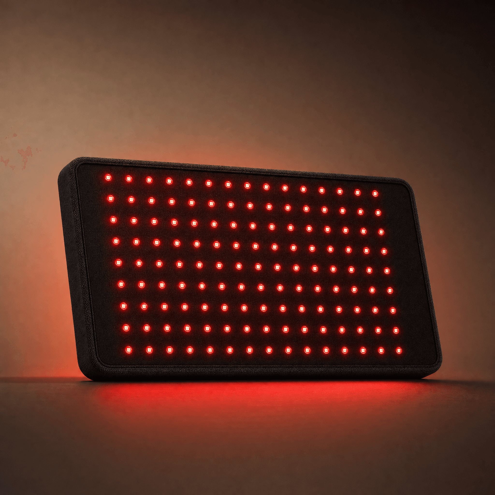
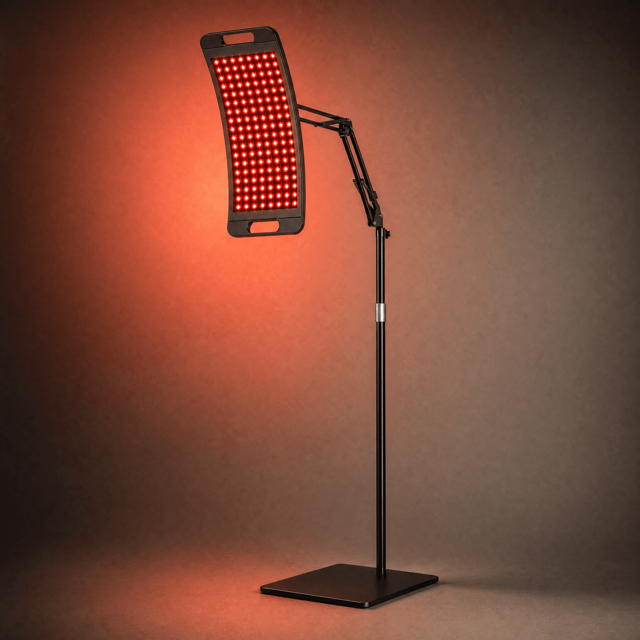
When you switch a Lumora device on, the panel emits two specific wavelengths of light: 660 nanometres (visible red) and 850 nanometres (near-infrared, invisible to the eye). These wavelengths sit in what researchers call the optical window of human tissue, the narrow band where light actually penetrates skin rather than bouncing off or being absorbed at the surface.
The mechanism is called photobiomodulation, or PBM. It was first identified in the 1960s by Endre Mester, a Hungarian physician who noticed that low-level laser exposure accelerated wound healing in mice. Six decades of peer-reviewed research later, PBM is one of the most studied modalities in regenerative medicine, with applications spanning skin, muscle, joint and bone recovery.
This page is a plain-English summary of what the research actually says.
How it works
Light absorbed by your mitochondria.
Every cell in your body contains hundreds to thousands of mitochondria, the organelles that produce adenosine triphosphate (ATP), the molecular fuel for nearly every process in human physiology.
Inside the mitochondrion, an enzyme called cytochrome c oxidase sits at the end of the electron transport chain. Cytochrome c oxidase is the primary photoacceptor for red and near-infrared light. When photons in the 600 to 1000 nm range hit it, a cascade follows: ATP synthesis increases, reactive oxygen species are modulated, and nitric oxide releases from the enzyme. Local circulation relaxes and the bottleneck that often slows recovery eases up.[8]
Why two wavelengths? 660 nm (red) is the workhorse for skin-level recovery; it is absorbed by surface tissue effectively. 850 nm (near-infrared) reaches deeper structures: muscle, joint capsule and the periosteum on the surface of bone, at clinically useful doses. Lumora devices fire both at the same time, so a single ten-minute session covers superficial and deep tissue at once.[11]
Focus 1 · Skin
The dermis is the most studied target for red light.
Collagen production
Brighter tone
Scar softening
Post-procedure recovery
The dermis sits closest to the light source and the results are visible, which is why most early photobiomodulation trials focused here. Three mechanisms are repeatedly demonstrated in the literature: fibroblast stimulation (the cells that make collagen and elastin), modulation of inflammation, and accelerated wound closure.[2]
A controlled trial published in Photomedicine and Laser Surgery measured intradermal collagen density via ultrasonography after 30 sessions of red and near-infrared light treatment. Treated subjects showed significant increases in collagen density and reductions in wrinkle depth compared to the control group.[1]
Photobiomodulation is now a routine adjunct to dermatological procedures (microneedling, laser resurfacing, post-surgical) to shorten the redness and downtime window. It is also used for acne, rosacea and post-inflammatory hyperpigmentation in clinical settings.
What this means in practice
Most users report subtle skin-tone improvements within four to six weeks of daily ten-minute sessions, with texture and elasticity changes continuing to compound through month two and three.
Built for this
Focus 2 · Muscle
Faster training rebound, less DOMS, quicker return to baseline.
ATP production up
Less DOMS
Faster rebound
Lower damage markers
For muscle, the research interest is recovery from exertion and injury. 850 nm penetrates skeletal muscle at clinically relevant doses, increasing local ATP availability and reducing oxidative stress in fibres damaged by training or strain. This translates to faster clearance of metabolic waste and less delayed-onset muscle soreness (DOMS).[4]
A 2015 systematic review and meta-analysis in Lasers in Medical Science pooled data from 39 randomised controlled trials covering phototherapy applied before or after exercise. Pooled effects favoured photobiomodulation for performance recovery, with lower creatine kinase markers (an indicator of muscle damage) and reduced perceived soreness in treated groups.[3]
The mechanism is consistent across athletic, occupational and rehabilitative contexts: more available ATP in damaged tissue, less oxidative stress, faster repair. The same engine that helps a marathon runner rebound also helps an office worker undo a long day at a desk.
What this means in practice
Athletes report faster training rebound and tighter cycles between hard sessions. Desk workers report less neck and back tightness in the evenings. Users recovering from soft-tissue strains report shorter total recovery windows.
Built for this
Focus 3 · Injury
Inflammation, joint pain, post-surgical recovery.
Joint pain reduced
Anti-inflammatory
Tendinopathy support
Post-surgical recovery
Injury here covers two overlapping research areas: chronic joint inflammation (osteoarthritis, tendinopathy, recurring pain) and acute soft-tissue injury or post-surgical recovery.
In addition to its ATP effects, photobiomodulation is anti-inflammatory. A 2017 review in AIMS Biophysics summarised the pathways: PBM modulates nuclear factor kappa B (NF-κB) and several inflammatory cytokines, dampening the inflammatory response without suppressing it. It is one of the few modalities that does this without pharmaceutical intervention.[7]
A 2003 systematic review by Bjordal and colleagues in the Australian Journal of Physiotherapy analysed location-specific dosing for chronic joint conditions and found significant pain reduction at appropriate doses in osteoarthritis, rheumatoid arthritis and chronic tendinopathy.[5] A Cochrane review the following year examined Class I, II and III laser therapy specifically for osteoarthritis and found moderate-quality evidence for short-term pain relief and improved function.[6]
What this means in practice
Users with recurring knee, shoulder or back pain typically report a reduction in flare frequency and severity over weeks of consistent use, particularly when sessions are timed near typical onset.
Built for this
Focus 4 · Bone
Stimulating the cells that build new bone.
Osteoblast activity
Bone matrix support
Density support
Fracture recovery
Bone is the most recent of the four research areas, with the strongest signals emerging since the mid-2000s. The mechanism: 850 nm penetrates to the periosteum and into trabecular bone at sufficient intensities, where it appears to stimulate osteoblast activity, the cells that build new bone matrix, while modulating osteoclast activity (the cells that break it down). The net tilt is toward bone formation.
An early animal study in the Journal of Photochemistry and Photobiology B irradiated rat tibia defects with low-power laser and found accelerated trabecular bone formation and improved histomorphometric markers compared to controls.[10] A 2006 review in Photomedicine and Laser Surgery by Pinheiro and Gerbi covered bone-specific applications including fracture healing, osseointegration around dental implants, and recovery from orthopaedic surgery.[9]
Human research is still expanding, but the early signals support the use of photobiomodulation as an adjunct in fracture recovery and as a supportive modality for users managing bone density concerns in mid-life and beyond.
What this means in practice
Most relevant for users managing post-fracture recovery, dental implant healing, or ongoing bone density support in mid-life. Bone effects are slower to register subjectively than skin or muscle, but the underlying mechanism is well-supported.
Built for this

Dosage & safety
The right dose, timed to ten minutes.
The clinical effect of red light depends on dose: how much energy per unit area, delivered over how long. Lumora devices are engineered to deliver doses within the “biphasic” response window identified in the literature. Enough to trigger cellular response, not so much that you overshoot into inhibitory effects.
Irradiance. Matched to evidence-based protocols (100+ mW/cm² at 6 inches on Aurora).
Session duration. Fixed at ten minutes via the built-in timer on every device.
Wavelengths. 660 nm + 850 nm, fired together. The most studied combination.
Red and near-infrared light is non-ionising and has no known cumulative damage profile at the doses Lumora devices deliver. Eye protection is provided in every box because direct retinal exposure to high-irradiance LED sources is uncomfortable and not recommended.
We recommend speaking to your GP before starting any new wellness routine, particularly if you are pregnant, taking photosensitising medication, or have a personal history of skin cancer.
References
The full reading list.
Every claim on this page is anchored to a peer-reviewed study. The full citations are below, numbered to match the inline markers.
- 1
Wunsch A, Matuschka K (2014). A controlled trial to determine the efficacy of red and near-infrared light treatment in patient satisfaction, reduction of fine lines, wrinkles, skin roughness, and intradermal collagen density increase.
Photomedicine and Laser Surgery. Photomed Laser Surg. 2014;32(2):93-100.
- 2
Avci P, Gupta A, Sadasivam M, Vecchio D, Pam Z, Pam N, Hamblin MR (2013). Low-level laser (light) therapy (LLLT) in skin: stimulating, healing, restoring.
Seminars in Cutaneous Medicine and Surgery. Semin Cutan Med Surg. 2013;32(1):41-52.
- 3
Leal-Junior EC, Vanin AA, Miranda EF, de Carvalho Pde T, Dal Corso S, Bjordal JM (2015). Effect of phototherapy (low-level laser therapy and light-emitting diode therapy) on exercise performance and markers of exercise recovery: a systematic review with meta-analysis.
Lasers in Medical Science. Lasers Med Sci. 2015;30(2):925-39.
- 4
Ferraresi C, Hamblin MR, Parizotto NA (2012). Low-level laser (light) therapy (LLLT) on muscle tissue: performance, fatigue and repair benefited by the power of light.
Photonics & Lasers in Medicine. Photonics Lasers Med. 2012;1(4):267-286.
- 5
Bjordal JM, Couppé C, Chow RT, Tunér J, Ljunggren EA (2003). A systematic review of low level laser therapy with location-specific doses for pain from chronic joint disorders.
Australian Journal of Physiotherapy. Aust J Physiother. 2003;49(2):107-116.
- 6
Brosseau L, Welch V, Wells G, et al. (2004). Low level laser therapy (Classes I, II and III) for treating osteoarthritis.
Cochrane Database of Systematic Reviews. Cochrane Database Syst Rev. 2004;(3):CD002046.
- 7
Hamblin MR (2017). Mechanisms and applications of the anti-inflammatory effects of photobiomodulation.
AIMS Biophysics. AIMS Biophys. 2017;4(3):337-361.
- 8
Karu TI (2010). Multiple roles of cytochrome c oxidase in mammalian cells under action of red and IR-A radiation.
IUBMB Life. IUBMB Life. 2010;62(8):607-610.
- 9
Pinheiro AL, Gerbi ME (2006). Photoengineering of bone repair processes.
Photomedicine and Laser Surgery. Photomed Laser Surg. 2006;24(2):169-178.
- 10
Garavello-Freitas I, Baranauskas V, Joazeiro PP, Padovani CR, Dal Pai-Silva M, da Cruz-Höfling MA (2003). Low-power laser irradiation improves histomorphometrical parameters and bone matrix organization during tibia wound healing in rats.
Journal of Photochemistry and Photobiology B: Biology. J Photochem Photobiol B. 2003;70(2):81-89.
- 11
de Freitas LF, Hamblin MR (2016). Proposed mechanisms of photobiomodulation or low-level light therapy.
IEEE Journal of Selected Topics in Quantum Electronics. IEEE J Sel Top Quantum Electron. 2016;22(3):348-364.
Built on this research
The dose, the wavelengths, the daily ritual.
Every Lumora device is engineered against the protocols cited above. Pick the form factor that fits your routine; the science is identical underneath.